Carotid artery exhibit for a wrongful death case: a model that shows where the measurement was taken
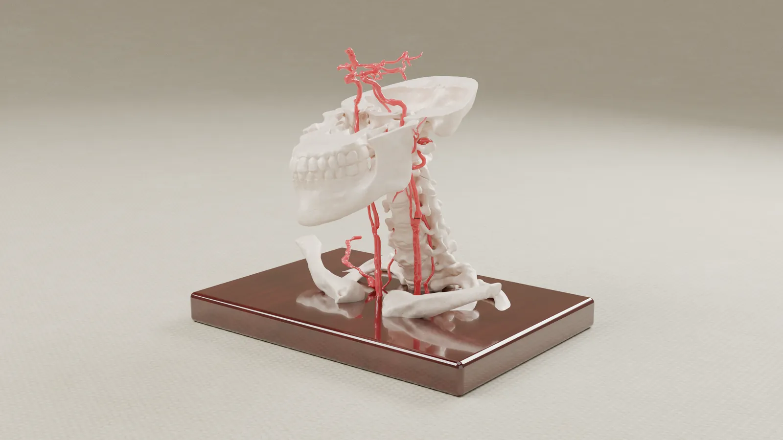
In a wrongful death case, the dispute turned on a CT measurement of the carotid artery and on where along the vessel it was taken. A CT slice looks exact, but the artery's width changes along its length, and a flat slice does not show where on the vessel it sits. The firm asked us for a model of the artery, from the aortic arch to the Circle of Willis, with the measured section marked.
- Practice area
- Wrongful death, medical malpractice
- Anatomy
- Carotid arteries from the aortic arch to the Circle of Willis, with the jaw, cervical spine and clavicles
- Client
- Plaintiff-side medical-malpractice firm
- Exhibits produced
- Artery model with the measured section marked, an enlarged artery section, interactive exhibit, renderings
The challenge
The width of the artery changes along its length, and a measurement means little unless it is tied to a place on the vessel. On a flat CT slice that place is hard to see. Counsel needed a jury to see the whole artery and the section where the measurement was taken.
The solution
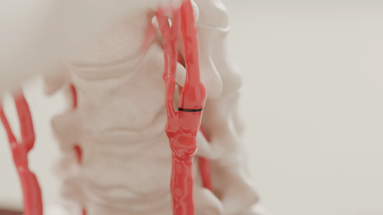
We built the arteries from the patient's own CT angiogram, from the aortic arch to the Circle of Willis, and added the jaw, the cervical spine and the clavicles for orientation. The section where the measurement was taken is marked in black, with the same artery visible above and below it. A second piece shows a section of the artery at twice the size.
A consulting radiologist reviewed the artery segmentation before we built the model. Counsel approved a digital proof before we built the final model.
We also built an interactive exhibit, in which a viewer can rotate the model and zoom to the measured slice.
What we built
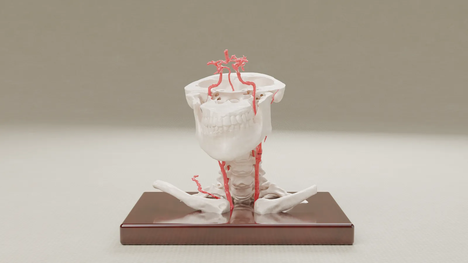
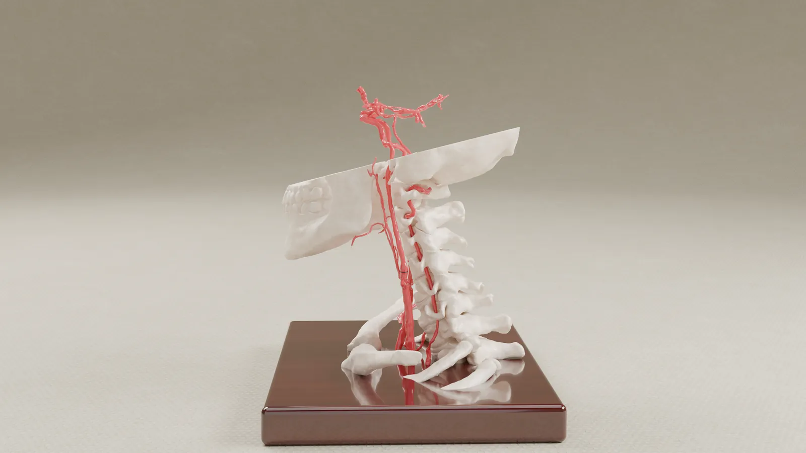
Artery model. A desktop model, about 7 by 5 by 8.5 inches, of the carotid arteries from the aortic arch to the Circle of Willis, with the jaw, cervical spine and clavicles on a wood display base. The measured section is marked in black.
Enlarged section. A section of the artery at twice the size, which makes the width at the measured section easier to see from a distance.
Interactive exhibit. A version on screen that a viewer can rotate and zoom to the measured slice.
Renderings. Views of the model on a display stand, and a close view of the marked section.
How it was built
- From the patient's own CT angiogram.
- A consulting radiologist reviewed the artery segmentation before the model was built.
- Counsel approved a digital proof before the final model was built.
- Built by the biomedical engineer (MS) you work with directly.
How it was used
The exhibit was delivered to counsel.
If your case turns on a measurement taken from a scan, send us the imaging and we will tell you what we would build.
Prior results do not guarantee a similar outcome. Case details are published with client permission; some details are anonymized to protect confidentiality. Every image on this page is a rendering of the exhibit.